Abdomen Anatomy Female Bowel : Large Intestine Wikipedia / The jejunum has the most developed and highest concentration.. It is approximately 5 meters long and includes the duodenum, jejunum, and ileum. However, the small intestine is present at the central part and lower parts of the abdominal proper cavity where the large intestine surrounds it. The ascending colon is the beginning of the large intestine into which the small intestine empties; The liver, stomach, and abdominal contents are clearly identified and labeled, including the cecum, ascending colon, transverse colon, descending colon, and small intestine. In women the lowest portion of the abdomen is actually the pelvis and involves the uterus fallopian tubes and ovaries.
• the descending colon travels down the left abdomen. We're going to take apart a plastic anatomy model and see what we can find in the abdomen. We'll identify as many organs as we can, see how they fit into the. The small intestine extends from the pylorus of the stomach, curling through the upper left and lower right quadrants of the abdomen. The small bowel contains prominent mucosal folds known as plicae circulares or valvular connvinetes.
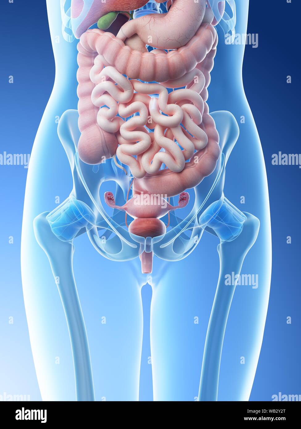
Anatomy Of The Female Abdomen And Pelvis Stock Photo Alamy from c8.alamy.com The organs of the digestive tract consist of the small and large intestines, the stomach, cecum, and the appendix.the stomach is located between the esophagus and the small intestine in the upper left region of the abdomen. Abdominal adhesions are the most common cause of obstruction of the small intestine. The photo of stomach and large intestine is on the woman's body against gray background, people with stomach ache problem concept, female anatomy intestines black icon, vector sign on isolated background. The small intestine is situated between the stomach and. The jejunum has the most developed and highest concentration. The major organs of the abdomen include the. However, the small intestine is present at the central part and lower parts of the abdominal proper cavity where the large intestine surrounds it. This medical exhibit diagram illustrates the anatomy of the female abdomen and pelvis from an anterior front cut away view showing elements of the digestive system the liver stomach and abdominal contents are clearly identified and labeled including the cecum ascending colon transverse colon descending colon and small intestine the image also shows the pelvis uterus and urinary bladder the pelvic bones and femur bones are ghosted beneath the skin
• the transverse colon runs across the abdomen.
It is designed to help the body absorb nutrients and fluids from the foods we eat and drink. National library of medicine was used as the basis to build an exemplary model of the female abdomen analyzing the normal anatomy we found several variations and pathologies of the vhf, such as missing muscles (gemellus superior, psoas. Various folds or reflections of the peritoneum connect viscera to the abdominal walls or to one another. It is in fact very difficult to identify the cause of an abdominal pain because of the multiple organs within. However, the small intestine is present at the central part and lower parts of the abdominal proper cavity where the large intestine surrounds it. As you can see in the below … This medical exhibit diagram illustrates the anatomy of the female abdomen and pelvis from an anterior front cut away view showing elements of the digestive system the liver stomach and abdominal contents are clearly identified and labeled including the cecum ascending colon transverse colon descending colon and small intestine the image also shows the pelvis uterus and urinary bladder the pelvic bones and femur bones are ghosted beneath the skin Abdominal adhesions are the most common cause of obstruction of the small intestine. It is about 6 m in length and extends from the pyloric sphincter to the ileocecal junction. Labeled structures include the large bowel (colon or large intestine), umbilicus, small intestine, ovary, fallopian tube, uterus and bladder. Portions of the small intestine, ascending colon, and right ureter may also extend into the rlq. • the transverse colon runs across the abdomen. We'll identify as many organs as we can, see how they fit into the.
This medical exhibit diagram illustrates the anatomy of the female abdomen and pelvis from an anterior front cut away view showing elements of the digestive system. • the sigmoid colon is a short curving of the colon, just before the rectum. The human intestine consists of two segments. This medical exhibit diagram illustrates the anatomy of the female abdomen and pelvis from an anterior front cut away view showing elements of the digestive system the liver stomach and abdominal contents are clearly identified and labeled including the cecum ascending colon transverse colon descending colon and small intestine the image also shows the pelvis uterus and urinary bladder the pelvic bones and femur bones are ghosted beneath the skin As you can see in the below …
Anatomy Of Stomach Area Koibana Info Anatomie Des Organes Anatomie Corps Humain Anatomie Du Corps from i.pinimg.com As you can see in the below … The right ovary and fallopian tube are also located in the right lower quadrant in females. However, the small intestine is present at the central part and lower parts of the abdominal proper cavity where the large intestine surrounds it. We're going to take apart a plastic anatomy model and see what we can find in the abdomen. This medical exhibit diagram illustrates the anatomy of the female abdomen and pelvis from an anterior front cut away view showing elements of the digestive system. The small intestine is situated between the stomach and. Abdominal adhesions are the most common cause of obstruction of the small intestine. Portions of the small intestine, ascending colon, and right ureter may also extend into the rlq.
1 the duodenum can be separated into.
The small intestine accomplishes this via a complex network of blood vessels, nerves, and muscles that wor … After taking out everything the body needs, the bowel then expels the leftover waste. It begins on the lower right side of the abdomen and then leads up to the transverse colon. Trial exhibits is a nationwide litigation support company with over 30 years of experience and involvement in thousands of jury trials, mediations, and arbitrations. Outline • abdominal cavity • external and internal fascia • abdominal muscle • intraperitoneal and retroperitoneal organs • ligaments • topography • laparotomy and its sites • summary However, the small intestine is present at the central part and lower parts of the abdominal proper cavity where the large intestine surrounds it. It is about 6 m in length and extends from the pyloric sphincter to the ileocecal junction. In latin, duodenum translates to 12 fingers, which is the approximate length of the organ. The organs of the digestive tract consist of the small and large intestines, the stomach, cecum, and the appendix.the stomach is located between the esophagus and the small intestine in the upper left region of the abdomen. This medical exhibit diagram illustrates the anatomy of the female abdomen and pelvis from an anterior front cut away view showing elements of the digestive system the liver stomach and abdominal contents are clearly identified and labeled including the cecum ascending colon transverse colon descending colon and small intestine the image also shows the pelvis uterus and urinary bladder the pelvic bones and femur bones are ghosted beneath the skin The ascending colon is the beginning of the large intestine into which the small intestine empties; At the level of the pelvic bones, the abdomen ends and the pelvis begins. The photo of stomach and large intestine is on the woman's body against gray background, people with stomach ache problem concept, female anatomy intestines black icon, vector sign on isolated background.
The beginning of the bowel is the small intestine, sometimes referred to as the small. The organs of the digestive tract consist of the small and large intestines, the stomach, cecum, and the appendix.the stomach is located between the esophagus and the small intestine in the upper left region of the abdomen. Portions of the small intestine, ascending colon, and right ureter may also extend into the rlq. The small bowel contains prominent mucosal folds known as plicae circulares or valvular connvinetes. • the transverse colon runs across the abdomen.
Small Intestine Digestive Disorders Merck Manuals Consumer Version from www.merckmanuals.com The colon is the longest part of the large intestine. At the level of the pelvic bones, the abdomen ends and the pelvis begins. However, the small intestine is present at the central part and lower parts of the abdominal proper cavity where the large intestine surrounds it. Abdominal adhesions can cause intestinal obstruction and female infertility. The diaphragm forms the upper surface of the abdomen. The jejunum has the most developed and highest concentration. The beginning of the bowel is the small intestine, sometimes referred to as the small. This medical exhibit diagram illustrates the anatomy of the female abdomen and pelvis from an anterior front cut away view showing elements of the digestive system the liver stomach and abdominal contents are clearly identified and labeled including the cecum ascending colon transverse colon descending colon and small intestine the image also shows the pelvis uterus and urinary bladder the pelvic bones and femur bones are ghosted beneath the skin
This medical exhibit diagram illustrates the anatomy of the female abdomen and pelvis from an anterior front cut away view showing elements of the digestive system.
The small intestine is a convoluted tube connecting the stomach with the large intestine. • the sigmoid colon is a short curving of the colon, just before the rectum. It is approximately 5 meters long and includes the duodenum, jejunum, and ileum. The colon is the longest part of the large intestine. Abdominal computed tomography (ct) is a type of medical imaging procedure used to diagnose and monitor internal stomach issues, like cancer, bowel obstruction, and abdominal pain. Portions of the small intestine, ascending colon, and right ureter may also extend into the rlq. It is about 6 meters (20 feet) long and extends from the pylorus of the stomach to the ileocecal junction. At the level of the pelvic bones, the abdomen ends and the pelvis begins. The liver stomach and abdominal contents are clearly identified and labeled including the cecum ascending colon transverse colon descending colon and small. Exhibit features the orientation of normal healthy colon anatomy within the female body along with a coronal section detailing some internal anatomy of the colon. National library of medicine was used as the basis to build an exemplary model of the female abdomen analyzing the normal anatomy we found several variations and pathologies of the vhf, such as missing muscles (gemellus superior, psoas. Various folds or reflections of the peritoneum connect viscera to the abdominal walls or to one another. Trial exhibits is a nationwide litigation support company with over 30 years of experience and involvement in thousands of jury trials, mediations, and arbitrations.
The photo of stomach and large intestine is on the woman's body against gray background, people with stomach ache problem concept, female anatomy intestines black icon, vector sign on isolated background abdomen anatomy-female. Intestinal obstruction is the partial or complete blockage of the movement of food, fluids, air, or stool through the intestines.


0 Komentar