Loculated Pleural Effusion Ct : Acquired Lesions Of The Lung And Pleura Clinical Gate / Both computed tomography (ct) and ultrasound (us) can be used to differentiate ascites from pleural effusion.
Loculated Pleural Effusion Ct : Acquired Lesions Of The Lung And Pleura Clinical Gate / Both computed tomography (ct) and ultrasound (us) can be used to differentiate ascites from pleural effusion.. Pleural fluid/serum ldh ratio >0.6. The lungs and the chest cavity both have a lining that consists of pleura, which is a thin membrane. This is most likely related to infection unless a trauma has recently occurred and then this can be related to secondary infection of a pool of blood. Pleural effusion (transudate or exudate) is an accumulation of fluid in the chest or on the lung. In healthy lungs, these membranes ensure that a.
Pleural effusions were measured by assessing the maximum perpendicular diameter to the parietal pleura at the greatest depth on axial ct images. Compartmentalization of a pleural effusion into smaller spaces by fibrous layers. Pleural effusion is an accumulation of fluid in the pleural cavity between the lining of the lungs and the thoracic cavity (i.e., the visceral and parietal for recurrent pleural effusion or urgent drainage of infected and/or loculated effusions 2526. Bilateral, left greater than right, pleural effusions with adjacent atelectasis and collapse versus consolidation of the left lower lobe. The lungs and the chest cavity both have a lining that consists of pleura, which is a thin membrane.
Figure 19 Imaging Of Pneumonia An Overview Springerlink from media.springernature.com It is important to assess both the quantity of the pleural effusion and severity of the atelectasis. Pleural infection pleural inflammation pleural malignancy (most often pleural fluid analysis findings: Pleural effusion is a condition in which excess fluid builds around the lung. Suspected parenchymal or pleural pathology. Pleural effusion can be a sign of serious illness. Pleural fluid/serum protein ratio >0.5. Pleural effusions represent a disturbance between pleural fluid production loculated pleural effusions: This is most likely related to infection unless a trauma has recently occurred and then this can be related to secondary infection of a pool of blood.
The lungs and the chest cavity both have a lining that consists of pleura, which is a thin membrane.
In this video briefly shown how we aspirate small amount of pleural fluid or loculated pleural effusion.for more videos please subscribe the channel.if you. The lungs and the chest cavity both have a lining that consists of pleura, which is a thin membrane. Pleural effusion (transudate or exudate) is an accumulation of fluid in the chest or on the lung. Ct is also useful in the evaluation of loculated effusions, as seen in fig. Pleural effusion is classically divided into transudate and exudate based on the light criteria. Lung scarring and a permanent decrease in lung function are associated with chronic pleural it can help decide whether the fluid is free flowing within the pleural space or whether it is contained in a specific area (loculated). Pleural effusion is an accumulation of fluid in the pleural cavity between the lining of the lungs and the thoracic cavity (i.e., the visceral and parietal ple… directed thoracentesis of a loculated effusion. The fluid is similar to water in its attenuation. Needle biopsy of the pleura can be done when thoracoscopy is unavailable. Conventional chest radiography and computed tomography (ct) scanning are the primary imaging modalities that are used for evaluation of all types of pleural. In the usa approximately 1.5 million people are diagnosed with a pleural effusion each year 2. Better quantification of the amount of fluid (compared. Pleural effusion due cardiovascular disease.
Pleural effusion is an abnormal, excessive collection of this fluid. Both computed tomography (ct) and ultrasound (us) can be used to differentiate ascites from pleural effusion. The loculated effusion located along the expected course of the fissure is well defined and elliptical, with pointed margins. Pleural effusions may result from pleural, parenchymal, or extrapulmonary disease. Pleural fluid ldh > two thirds of upper limit for serum ldh.
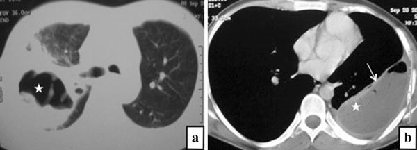
Pleural Effusion Loculation Ct Chest Page 1 Line 17qq Com from img.17qq.com Conventional chest radiography and computed tomography (ct) scanning are the primary imaging modalities that are used for evaluation of all types of pleural. Pleural effusions occur as a result of increased fluid formation and/or reduced fluid resorption. Both computed tomography (ct) and ultrasound (us) can be used to differentiate ascites from pleural effusion. Freely mobile pleural effusions are easily proven with decubitus chest films, but loculated subpulmonic effusions can mimic intraabdominal fluid. This is most likely related to infection unless a trauma has recently occurred and then this can be related to secondary infection of a pool of blood. Pleural fluid ldh > two thirds of upper limit for serum ldh. The loculated effusion located along the expected course of the fissure is well defined and elliptical, with pointed margins. Us scan they can be identified clearly and it is very complicated.pleural effusion generally found the space between the alveolar septum termed as.
Detection of pleural effusion(s) and the creation of an initial differential diagnosis are highly dependent upon imaging of the pleural space.
The pleura are thin membranes that line the lungs and the inside of the chest cavity and act to lubricate and facilitate breathing. Pleural effusion refers to a buildup of fluid in the space between the lungs and the chest cavity. The fluid is similar to water in its attenuation. Pleural effusions were measured by assessing the maximum perpendicular diameter to the parietal pleura at the greatest depth on axial ct images. Pleural effusion (fluid around the lungs) picture and facts. Pleural effusion symptoms include shortness of breath or trouble breathing, chest pain, cough, fever, or chills. Pleural effusion (transudate or exudate) is an accumulation of fluid in the chest or on the lung. The split pleura sign represents a rind of visceral and parietal pleural thickening surrounding a loculated effusion (figure 13). Pleural fluid ldh > two thirds of upper limit for serum ldh. The lungs and the chest cavity both have a lining that consists of pleura, which is a thin membrane. Send aspirated fluid for cytology. Both computed tomography (ct) and ultrasound (us) can be used to differentiate ascites from pleural effusion. Pleural effusions occur as a result of increased fluid formation and/or reduced fluid resorption.
Freely mobile pleural effusions are easily proven with decubitus chest films, but loculated subpulmonic effusions can mimic intraabdominal fluid. Radiography, ultrasound and chest ct reveal the presence of free or loculated pe, occasionally with images compatible with clots that may also reveal. Suspected parenchymal or pleural pathology. Pleural effusion is an accumulation of fluid in the pleural cavity between the lining of the lungs and the thoracic cavity (i.e., the visceral and parietal ple… directed thoracentesis of a loculated effusion. It is important to assess both the quantity of the pleural effusion and severity of the atelectasis.
Pleural Empyema Wikipedia from upload.wikimedia.org Pleural infection pleural inflammation pleural malignancy (most often pleural fluid analysis findings: Pleural fluid ldh > two thirds of upper limit for serum ldh. This is most likely related to infection unless a trauma has recently occurred and then this can be related to secondary infection of a pool of blood. Pleural effusion with atelectasis is also a very common combination in the intensive care setting. If none is present the fluid is virtually always a transudate. Bilateral, left greater than right, pleural effusions with adjacent atelectasis and collapse versus consolidation of the left lower lobe. Pleural effusion due cardiovascular disease. Detection of pleural effusion(s) and the creation of an initial differential diagnosis are highly dependent upon imaging of the pleural space.
Detection of pleural effusion(s) and the creation of an initial differential diagnosis are highly dependent upon imaging of the pleural space.
Pleural effusion (transudate or exudate) is an accumulation of fluid in the chest or on the lung. In healthy lungs, these membranes ensure that a. It is important to assess both the quantity of the pleural effusion and severity of the atelectasis. Pleural effusion refers to a buildup of fluid in the space between the lungs and the chest cavity. Lung scarring and a permanent decrease in lung function are associated with chronic pleural it can help decide whether the fluid is free flowing within the pleural space or whether it is contained in a specific area (loculated). Ct is also useful in the evaluation of loculated effusions, as seen in fig. Bilateral, left greater than right, pleural effusions with adjacent atelectasis and collapse versus consolidation of the left lower lobe. Although pleural effusions are often easily identified on computed tomography (ct), trace on ct, pleural thickening may be difficult to distinguish from an effusion. Pleural fluid/serum ldh ratio >0.6. The annual incidence of pleural effusion in the developed world has been estimated at 320 per 100,000 population per year 1. It can result from pneumonia and many other conditions. The split pleura sign represents a rind of visceral and parietal pleural thickening surrounding a loculated effusion (figure 13). Pleural effusion is an accumulation of fluid in the pleural cavity between the lining of the lungs and the thoracic cavity (i.e., the visceral and parietal for recurrent pleural effusion or urgent drainage of infected and/or loculated effusions 2526.
Pleural effusion due cardiovascular disease loculated pleural effusion. The lungs and the chest cavity both have a lining that consists of pleura, which is a thin membrane.


0 Komentar